Pseudomonas aeruginosa is a human pathogen that forms robust biofilms that extensively tolerate antibiotics and effectively evade clearance by the immune system. Two of the important bacterial-produced polymers in the matrices of P. aeruginosa biofilms are alginate and extracellular DNA (eDNA), both of which are anionic and therefore have the potential to interact electrostatically with cations. Many physiological sites of infection contain significant concentrations of the calcium ion (Ca2+). We study the structural and mechanical impacts of Ca2+ supplementation in alginate-dominated biofilms grown in vitro and we evaluate the impact of targeted enzyme treatments on clearance by immune cells. We use multiple particle tracking microrheology to evaluate the changes in biofilm viscoelasticity caused by treatment with alginate lyase and/or DNAse I. For biofilms grown without Ca2+, we have correlated a decrease in relative elasticity with increased phagocytic success. However, we have found that growth with Ca2+ supplementation disrupts this correlation except in the case where both enzymes are applied. This suggests that the calcium cation may be impacting the microstructure of the biofilm in non-trivial ways. Confocal laser scanning fluorescence microscopy and scanning electron microscopy reveal unique Ca2+-dependent eDNA and alginate microstructures. Our work suggests that the presence of Ca2+ drives the formation of structurally and compositionally discrete microdomains within the biofilm through electrostatic interactions with the anionic matrix components eDNA and alginate. We study how these structures serve a protective function and how to compromise them to render bacteria susceptible to phagocytosis.
Shown above is a biofilm whose mechanics have just been measured on a rheometer by Ph.D. students Kristin Kovach (Physics) and Megan Davis-Fields (Microbiology and Molecular Genetics).
Biofilms are complex communities of interacting microorganisms embedded in a matrix of polymer, protein, and other materials. Being in a biofilm gives microbes physical and chemical protection against antibiotic treatments and immune clearance. In clinical infections, the formation of biofilms presents a significant challenge, accounting for approximately 80% of chronic infections, and resulting in substantial financial burdens on the healthcare system and adversely affecting numerous lives.
In addition to self-production, the EPS components can also originate from incorporation of environmental materials. An example of this is the incorporation of collagen, a prevalent protein in infections, into the biofilm matrix, potentially causing modifications to its structural and mechanical properties.
Shown above is a micrograph movie of a human neutrophil (top) eating bacteria out of a biofilm. This movie was taken by Ph.D. student Layla Bakhtiari (Physics).
Phagocytosis is a mechanical mode of immune clearance that involves the engulfment and internalization of pathogens. In our previous work using much-larger-than-neutrophils gels as phagocytosis targets, we found that the success rate of and timescale required for successful phagocytosis depend on the elasticity and toughness of the gel target structure. Given the changes in biofilm mechanics arising from the incorporation of collagen, we hypothesize that the incorporation of collagen within biofilms may affect the efficacy of immune cells at phagocytosing biofilm bacteria.
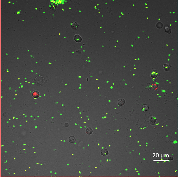
Picture of human neutrophils (greyscale) cultured with P. aeruginosa biofilm. The bacteria constitutively express Green Fluorescent Protein, and were stained with pHrodo Red succinimidyl ester, which emits red fluorescence in acidic phagosomes. Thus, the red bacteria have been phagocytosed. Image by Ph.D. student Marilyn Wells (Physics).
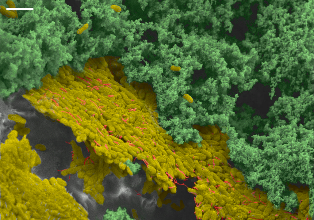
A scanning electron micrograph shows a dense “shield” of biofilm bacteria formed by the interaction of extracellular DNA and calcium. Image by Ph.D. student Marilyn Wells (Physics).