Associate Professor, University of Texas at Austin (2018-present)
Assistant Professor, University of Texas at Austin (2010-2018)
Postdoc, University of Illinois at Champaign-Urbana (2006-2010)
Postdoc, University of Edinburgh (2003-2006)
Education
Ph.D. in Physics Harvard University (2003)
B.Sc. in Physics and Mathematics Vanderbilt University (1997)
Awards and Honors
fellow on Elizabeth B. Gleeson Professorship in Plan II (Plan II Honors Program at the University of Texas at Austin) (2023)
Physics fellow, Texas Mindset Initiative, University of Texas, Austin (2023)
fellow on Trull Centennial Professorship in Physics, University of Texas, Austin (2022-2025)
Provost’s Mentored Faculty Fellow, University of Texas, Austin (2022 – 2023)
Provost’s Teaching Fellow, University of Texas, Austin (2020 – 2024)
Teaching Excellence Award, College of Natural Sciences, University of Texas, Austin (2020)
President’s Associates Teaching Excellence Award, University of Texas, Austin (2019)
Travel award from Rom Rhome International Professional Development Fund, University of Texas at Austin (2018)
Mitchell Award (2015) to Nalin Ratnayeke for outstanding undergraduate research done under my supervision
Hyer Research Award (2013) from the Texas Section of the American Physical Society. This award is for best undergraduate research and was given jointly to me and Nalin Ratnayeke.
Cystic Fibrosis Foundation Postdoctoral Fellow (2008-2010)
Magna Cum Laude & Honors in Physics, Vanderbilt University(1997)
We are actively recruiting PhD students
If you are a prospective Ph.D. student, please email Vernita: gordon at chaos dot edu. Tell her what about her group is interesting to you and how it fits into your background. If you don’t hear back from her, wait a week and try again. Sometimes she gets overwhelmed and doesn’t respond to emails, but that’s her fault, not yours. She still wants to hear from you.
Vernita can take Ph.D. students from the Physics, Microbiology and Molecular Genetics, Cell and Molecular Biology, and Biomedical Engineering programs. She thinks she is a good advisor and she encourages you to contact her current group members to find out more about what life in her group is like.
We also take undergraduate researchers. If you are interested in doing undergraduate research, email Vernita. Tell her about your background and why you’re interested in doing research in her group. If you don’t hear back from her, wait a week and try again. Sometimes she gets overwhelmed and doesn’t respond to emails, but that’s her fault, not yours. She still wants to hear from you.
More generally, the biophysical questions we ask address important topics because they address processes that are foundational to multicellular life (including ourselves and you, Dear Reader). We want especially to know how physics shapes these biological interactions and how physical tools and modes of thought can help us understand them better.
Motivated postdocs, graduate, undergraduate students will have the opportunity to contribute to multiple strands of fast-moving research within this theme. Our toolkit includes a variety of techniques for microscope imaging, micromanipulation, and computational analysis, and is likewise constantly expanding.
For more information on the group’s research and/or opportunities in the group, please email Vernita or come find us in person on the 14th floor of PMA.
Vernita’s Group
Our group focuses on understanding how physical characteristics, like mechanics and spatial structure, influence biology in complex multicellular systems, and vice versa – how do biological systems control their physical properties, and how does this benefit the biological system?
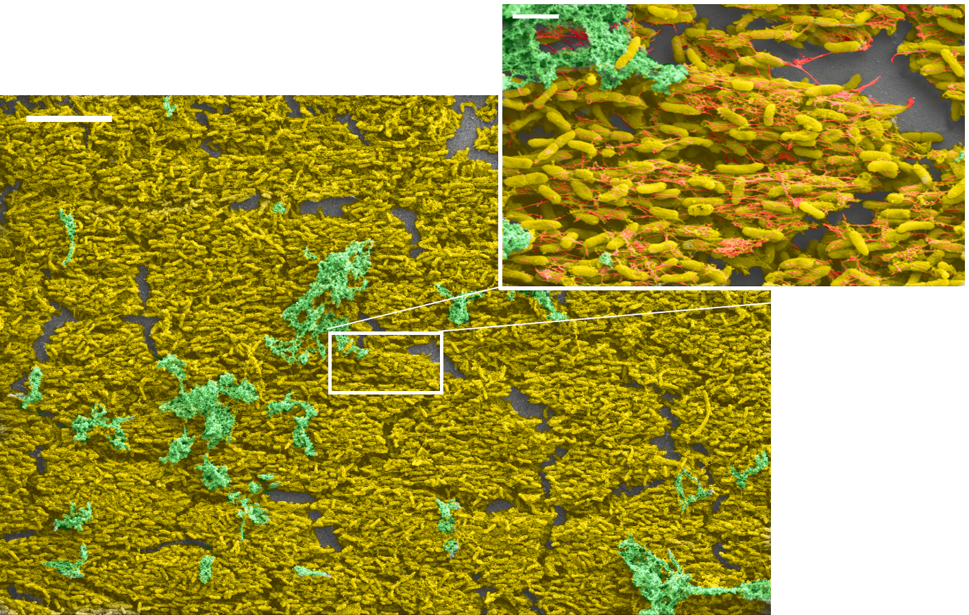
Marilyn Wells, Physics Ph.D. student, took these scanning electron microscope pictures of biofilms.
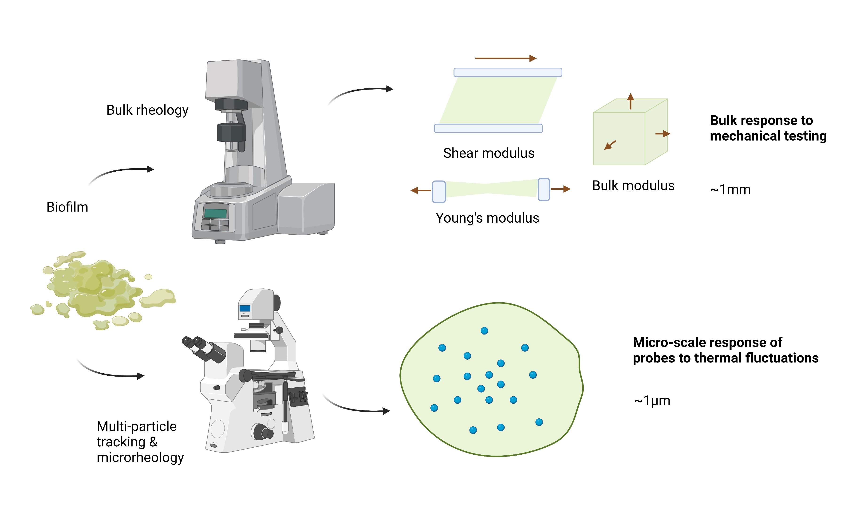
Our work focuses primarily on bacterial biofilms, which can cause harm in infection and in the built environment. We want to know how we could manipulate physical characteristics, of the biofilms and/or their environments, to prevent or ameliorate harmful biofilms. A biofilm can be considered as a heterogeneous soft multicomponent material, with viscoelastic mechanics and complex microstructure.
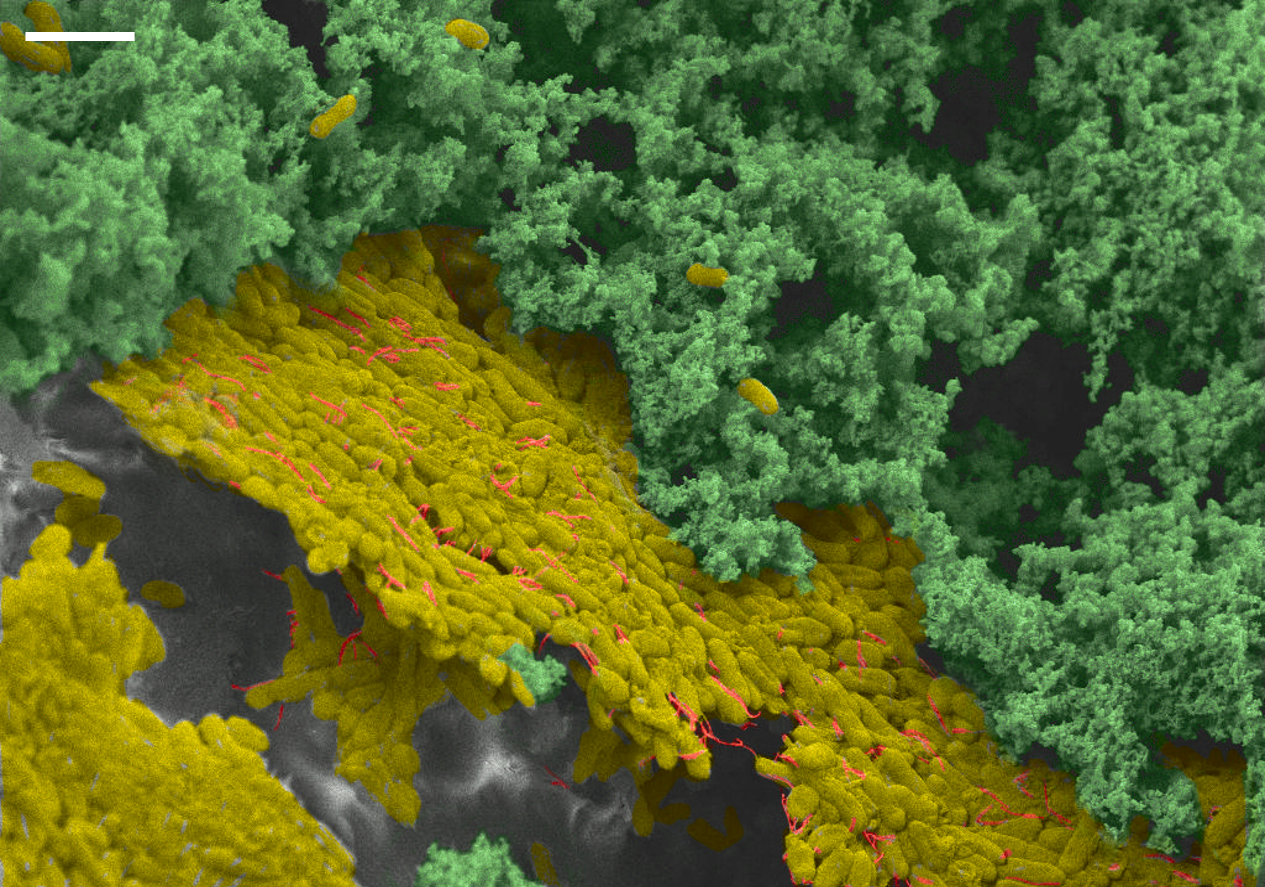
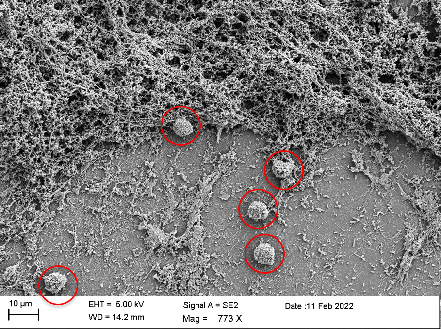
Marilyn Wells, Physics PhD student, took this scanning electron microscope picture of white blood cells interacting with a biofilm. White blood cells (circled in red) are an important component of the immune system.
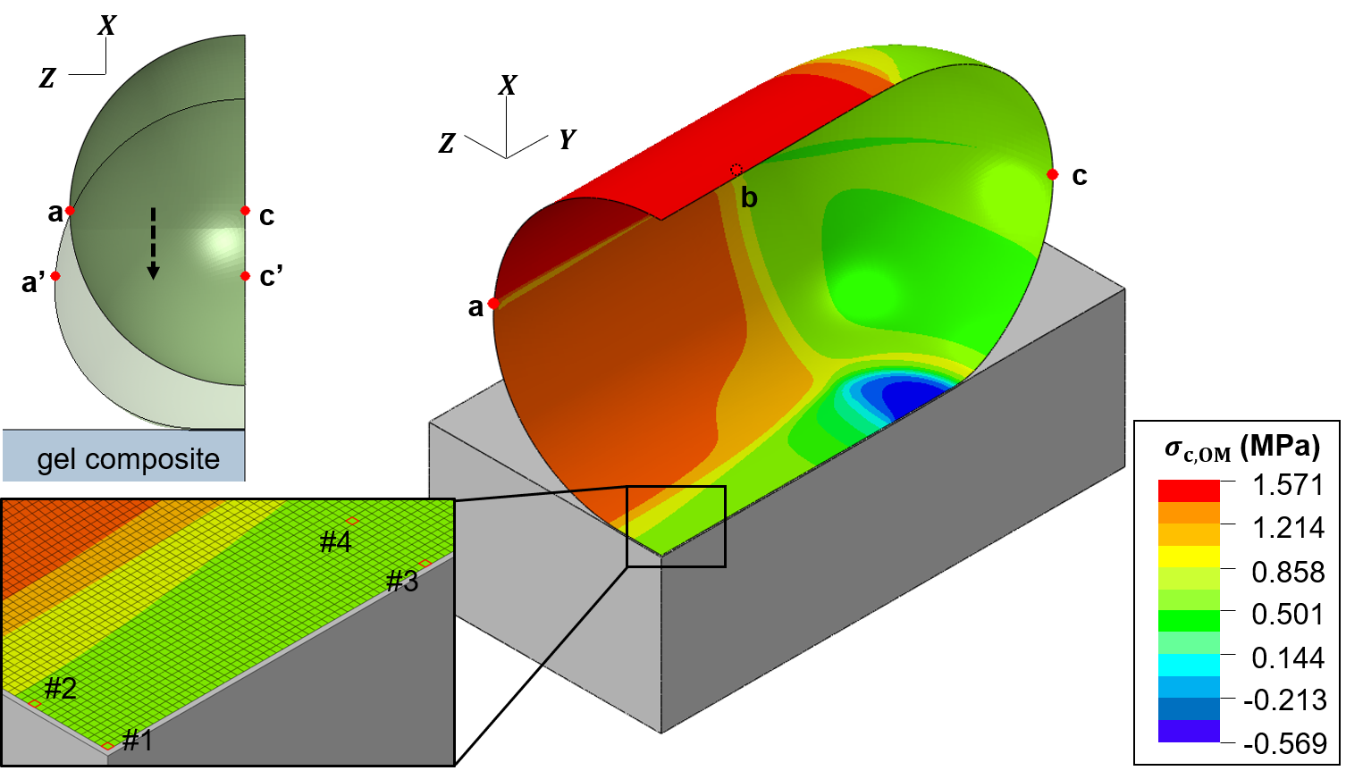
Chad Wong, Mechanical Engineering PhD student, did this finite-element modeling to describe the changes in mechanical stress and strain that a bacterial cell envelope experiences when it attaches to a surface, and how the changes in the bacterial cell envelope depend on the stiffness of the surface.
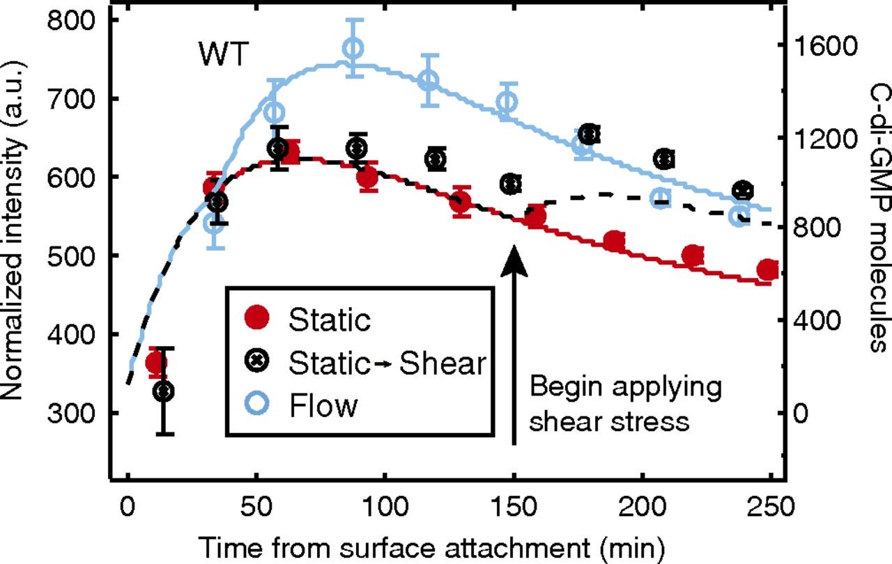
Chris Rodesney, Physics PhD student, did analytical modeling to describe his experimental measurements of bacterial signaling as a function of shear stress. Experimental data for three different stress timecourses are shown as discrete points of three different colors; modeling results for three different stress timecourses are shown as lines of the corresponding colors. https://www.pnas.org/doi/10.1073/pnas.1703255114
Bacteria start forming a biofilm when they encounter a surface or other interface. We are working to understand how interfacial interactions give rise to the biological signals that cause a biofilm to form. Our primary focus is on effects arising from surface stiffness and surface structure, and how bacterial mechanosensing of physical cues (such as material properties and forces) gives rise to an active biological response. We use modeling to gain insight into things that aren’t experimentally accessible, and to guide us in interpreting our experimental results. Here are some of our recent papers where we used these techniques: https://www.pnas.org/doi/10.1073/pnas.1703255114https://www.biorxiv.org/content/10.1101/2023.01.26.525810v1https://royalsocietypublishing.org/doi/10.1098/rsos.201453
Bacteria and Radiation
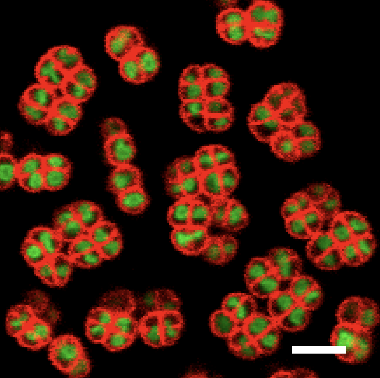
Brandon Niese, Physics PhD student, took these laser-scanning confocal microscope pictures of Deinococcus radiodurans.D. radiodurans is the most radiation-resistant bacteria known – the bacteria shown have been exposed to radiation levels up to 12 kGy, orders of magnitude more than what are needed to kill most bacterial and mammalian cells.
More recently, we have been turning our attention to the effects of radiation on bacteria. In one project, we are working with a group in Chemical Engineering (https://sites.utexas.edu/contreraslab/) to study how exposure to radiation and changes in protein expression is linked to changes in the size and internal structure of bacteria. We use machine learning and quantitative image analysis to measure the sizes and shapes of bacteria and their nucleoids.
With another group in Physics (https://raizenlab.ph.utexas.edu/), we are determining the role radioactive isotopes could play in creating self-sterilizing surfaces that prevent the development of biofilms.
Group Fun
Sometimes we go out for nachos.
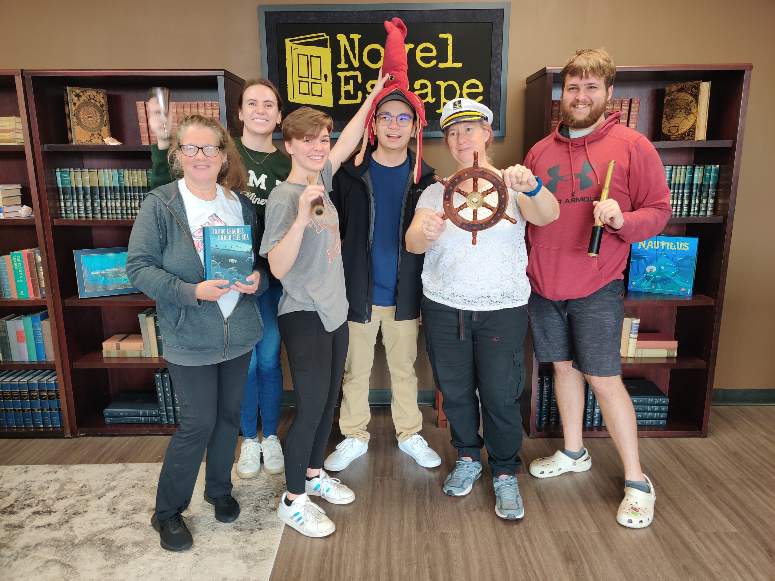
Sometimes we do an escape room and are victorious.

 The role of mechanics in biofilm formation
 Biofilm mechanics and the immune system
 Spatial structure and biofilms
 Outreach & Education
 Bacteria and radiation
 Brandon Niese
 Marilyn Wells
 Yu-Chern Chad Wong
 Xuening Zhou