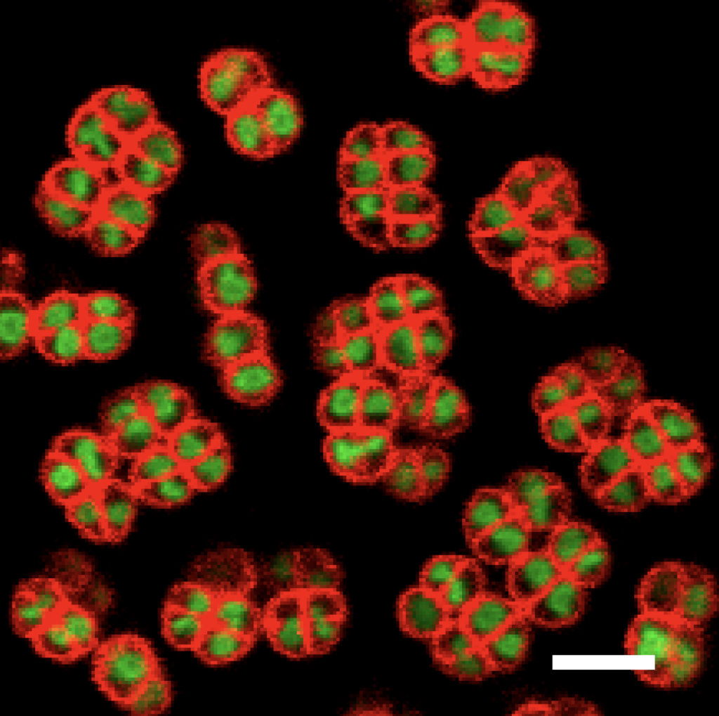
Cells of Deinococcus radiodurans have been imaged using confocal microscopy by Ph.D. student Brandon Niese (Physics).
Dienococcus radiodurans was discovered 1956 during experiments to determine whether canned food can be sterilized by using high doses of gamma radiation. A tin of meat was exposed but still spoiled, and thus D. radiodurans was isolated. In its more recent history, it was found to survive for 3 years in space. For all these conditions it can survive in, it is known as an “extremophile,” which means an organism that can survive and even sometimes thrive in environments that would kill most other organisms. According to the Guinness Book of World Records it is the “worlds toughest known bacterium.”
D. radiodurans has a spherical morphology and is about 1-2 microns in diameter. It divides along 2 perpendicular axis and thus groups in tetrads. Prokaryotes, like D. radiodurans, don’t have a membrane bound nucleus to hold their genetic material, so they have a compact mass of DNA (called the nucleoid). D.radiodurans have a relatively compact nucleoid compared to other bacteria of its size and when it becomes irradiated it becomes even more condensed. Previously work has shown that after irradiating D. radiodurans, the structure of the nucleoid changes and parts of the chromosome rearranges physical locations. In collaboration with the Contreras group in Chemical Engineering, which has extensive experience with D.radiodurans, we are developing tools for high-throughput microscopy and analysis, and using them to study nucleoid dynamics and condensation, and their dependence on specific proteins and environmental conditions.